Product Description
Product Description
Alpha-Pseudovirus for Bunyaviridae
The order of Bunyaviridae include the Nairoviruses, like Crimean-Congo Hemorrhagic Fever virus (CCHFV), Phleboviruses, such as Rift Valley Fever (RVFV) and the Hantaviruses like Andes virus (ANDV).
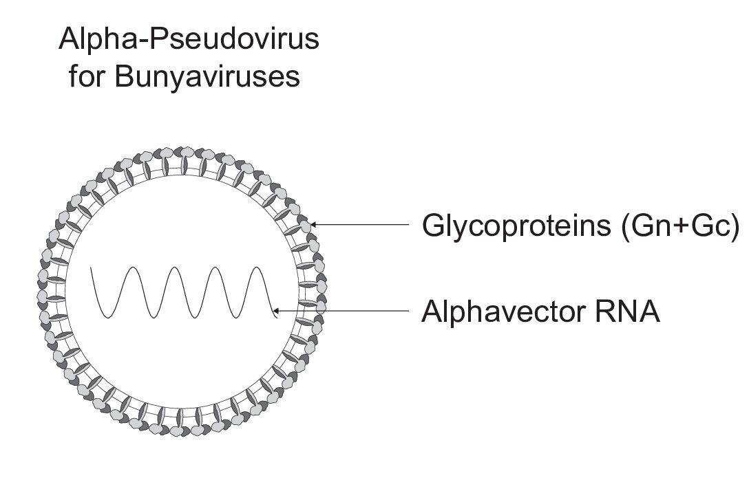
The rapid alpha-pseudoviruses for viruses belonging to the Bunyaviridae order are assembled with the glycoproteins Gn and Gc and package a self-amplifying RNA genome derived from an alphavirus vector. The Crimean-Congo Hemorrhagic Fever virus (CCHF) alpha-pseudovirus is based on NCBI reference sequenceADQ57289.1 . The Rift Valley Fever virus (RVFV) alpha-pseudovirus is based on NCBI reference sequence ABD38813.1. The Andes virus (ANDV) alpha-pseudovirus is based on the NCBI reference sequence NP_604472.1.
Applications:
- Facilitating ANDV, CCHFV, RVFV pseudovirus transduction of target cells for viral entry and functional studies
- Enabling rapid Anti-ANDV, Anti-CCHFV, and Anti-RVFV drug screening
- Supporting rapid Anti-ANDV, Anti-CCHFV, and Anti-RVFV neutralizing antibody screening
Description:
A novel rapid hybrid alpha-pseudovirus for Bunyaviruses including Crimean-Congo Hemorrhagic Fever virus (Ha-CCHFV), Rift Valley Fever virus (Ha-RVFV) and Andes virus (Ha-ANDV).
The alpha-pseudoviruses are single-cycle viruses with self-replicating RNA for rapid quantification of neutralizing antibodies and entry-inhibiting drugs. These pseudoviruses are BSL-2 safe and ready to use for studying viral entry. Additionally, we help our customers to assemble our hybrid alpha-pseudoviruses at any scale.
To enhance your pseudovirus entry, ask about receiving a free sample of our propriety InfectinTM which can significantly promote productive viral infection in a variety of host cells, enhancing viral infection rates by 3 to 20-fold.
Background:
Bunyaviruses constitute the largest group of RNA viruses, causing a spectrum of febrile and hemorrhagic illnesses worldwide. Originally identified as a single serotype in Africa, their number has now surpassed 500, with global distribution. These viruses, predominantly tri-segmented and single-stranded, are transmitted primarily by arthropod and rodent vectors, infecting various animals and plants. Despite a small number of encoded proteins, bunyaviruses can lead to potentially fatal diseases, adept at suppressing the host’s innate antiviral immune mechanisms. This review provides an overview of the Bunyavirales order, detailing their mechanisms of infection, replication, and evasion of host immune responses. Taxonomically reclassified as the only order of the class Ellioviricetes, Bunyavirales encompass fourteen families. Structurally, bunyaviruses consist of tri-segmented negative/ambisense RNA, with the Large (L), Medium (M), and Small (S) segments forming ribonucleoprotein structures complexed to nucleoproteins. These segments encode proteins crucial for viral replication and assembly, including the RNA-dependent RNA polymerase (RdRp) and glycoproteins Gn and Gc. Additionally, bunyaviruses may encode non-structural proteins (NSm and NSs), implicated in inhibiting the host immune response. Bunyaviruses enter the host cell via receptor-mediated endocytosis through the surface glycoprotein Gc, which is responsible for binding to the cellular receptors. Upon attachment, Gc responds to the reduced pH of endocytic compartments with a conformational change that results in a fusion loop, allowing it to insert into the endosomal membrane. Gc then folds back on itself, forcing the cell membrane (held by the fusion loop) and the viral membrane (held by a trans-membrane anchor) against each other, resulting in fusion and releasing the viral genome into the cytoplasm. Some of the receptors facilitating the viral entry that have been described include clathrin-dependent endocytosis, 3 integrins, nucleolin, and DC-SIGN (a C-type lectin restricted to interstitial dendric cells and certain tissue macrophages). Understanding bunyavirus structure, genomics, and immune response evasion is essential for developing effective control measures against these diverse and potentially deadly pathogens.
Crimean-Congo hemorrhagic fever (CCHF) is a lethal viral infection with global significance, primarily transmitted by ticks of the genus Hyalomma. The virus, CCHFV, belongs to the family Nairovirus within the Bunyaviridae order, with circulation dependent on tick distribution. Originating 3,100–3,500 years ago, it was first reported in Crimea in 1944, leading to its initial designation as Crimean hemorrhagic fever, later renamed to Crimean-Congo hemorrhagic fever after similar reports in the Congo in 1969. CCHFV exhibits a widespread geographical distribution, spanning Africa, Asia, Europe, and the Middle East, and has shown emergence in the Balkans since 2002. The virus is clustered into seven genotypes based on genetic variation. Highly pathogenic, easily transmissible, and with a high case-fatality rate of 10–40%, CCHFV poses a severe global health threat. In Pakistan, it has become an epidemic, necessitating urgent preventive measures. Due to its highly pathogenic nature, CCHFV is restricted to biosafety level four (BSL-4) laboratories. Structurally, CCHFV is spherical, with a lipid envelope and glycoprotein spikes, containing single-stranded RNA divided into three segments forming a ribo-nucleocapsid. Clinical symptoms include hemorrhage and a high case-fatality rate underscores the importance of understanding CCHFV’s virology, transmission pathways, and control measures to mitigate its impact on public health.
Rift Valley Fever Virus (RVFV), a member of the Bunyaviridae order, specifically the Phlebovirus family, is primarily transmitted by mosquitoes and poses a significant threat to both humans and animals. First identified in 1930 during an outbreak in Kenya’s Rift Valley, RVFV’s geographic distribution has expanded to include most countries in Africa, Madagascar, and regions beyond, such as the Arabian Peninsula and the Archipelago of Comores. Concerns about its potential introduction into RVF-free regions like Europe and the USA have escalated due to factors like climate change and international trade in live animals. RVFV’s genome consists of three segments, with replication occurring in the cytoplasm and virions maturing in the Golgi compartment. Recent studies have revealed an icosahedral symmetry in the virion structure, underscoring its complex packaging mechanism. Efforts to understand RVFV’s molecular epidemiology, genetics, vectors, diagnostics, and pathogenesis are crucial for effective surveillance and control of this emerging infectious disease, given its severe disease outbreaks in both animals and humans.
Andes virus (ANDV) is a deadly viruses belonging to the hantavirus familywithin the Bunyaviridae order. ANDV stands out among hantaviruses as a significant cause of hantavirus pulmonary syndrome (HPS) cases in Argentina, Chile, and Uruguay. While rodents are recognized as the primary reservoirs for hantaviruses, ANDV presents an additional route of transmission through person-to-person spread, a feature unique among hantaviruses. This transmission mode was first documented during an HPS outbreak in southwest Argentina in 1996, highlighting ANDV’s exceptional characteristics. ANDV encompasses six distinct lineages, each associated with specific regions in Argentina. While clusters of HPS cases are typically linked to rodent exposure, our analysis reveals new evidence of interhuman transmission, particularly with ANDV Sout lineage. Additionally, we report the first instance of another lineage, ANDV Cent BsAs, being implicated in person-to-person transmission. This transmission likely occurs during the prodromal phase or shortly after, facilitated by close and prolonged contact. Understanding these transmission dynamics is crucial for effective control and prevention strategies against this deadly virus.
If you have any additional questions please contact us by email: info@virongy.com
Example of results:
Rapid ANDV alpha-pseudovirus luciferase transduction of Vero cells (Left): Vero cells were transduced with Ha-ANDV(Luc) alpha-pseudovirus (with a luciferase reporter). Reporter expression was quantified at 24 hours post-transduction (luciferase assay).
Rapid ANDV alpha-pseudovirus GFP transduction of Vero cells (Right): Vero cells were transduced with Ha-ANDV (GFP) alpha-pseudovirus (with a GFP reporter). Reporter expression was quantified at 24 hours post-transduction (GFP flow cytometry).

Ha-CCHFV, Ha-RVFV and Ha-ANDV pseudoviruses are intended for Research Use Only and are not for diagnostic or therapeutic purposes or use in humans or animals.
Documents
Documents
References
References
Related links:
- Boshra H. An Overview of the Infectious Cycle of Bunyaviruses. Viruses. 2022; 14(10):2139. https://doi.org/10.3390/v14102139
- Aslam S, Latif MS, Daud M, Rahman ZU, Tabassum B, Riaz MS, Khan A, Tariq M, Husnain T. Crimean-Congo hemorrhagic fever: Risk factors and control measures for the infection abatement. Biomed Rep. 2016 Jan;4(1):15-20. doi: 10.3892/br.2015.545. Epub 2015 Nov 18. PMID: 26870327; PMCID: PMC4726894.
- Pepin M, Bouloy M, Bird BH, Kemp A, Paweska J. Rift Valley fever virus(Bunyaviridae: Phlebovirus): an update on pathogenesis, molecular epidemiology, vectors, diagnostics and prevention. Vet Res. 2010 Nov-Dec;41(6):61. doi: 10.1051/vetres/2010033. PMID: 21188836; PMCID: PMC2896810.


